Dark-field microscopy of blood from healthy individuals revealed the existence of pleomorphic microorganisms.
These bacteria exhibited limited growth and susceptibility to antibiotics and could be detected by fluorescent in situ hybridization and flow cytometry. They were further characterized by analysis of their 16S rRNA and gyrB genes.
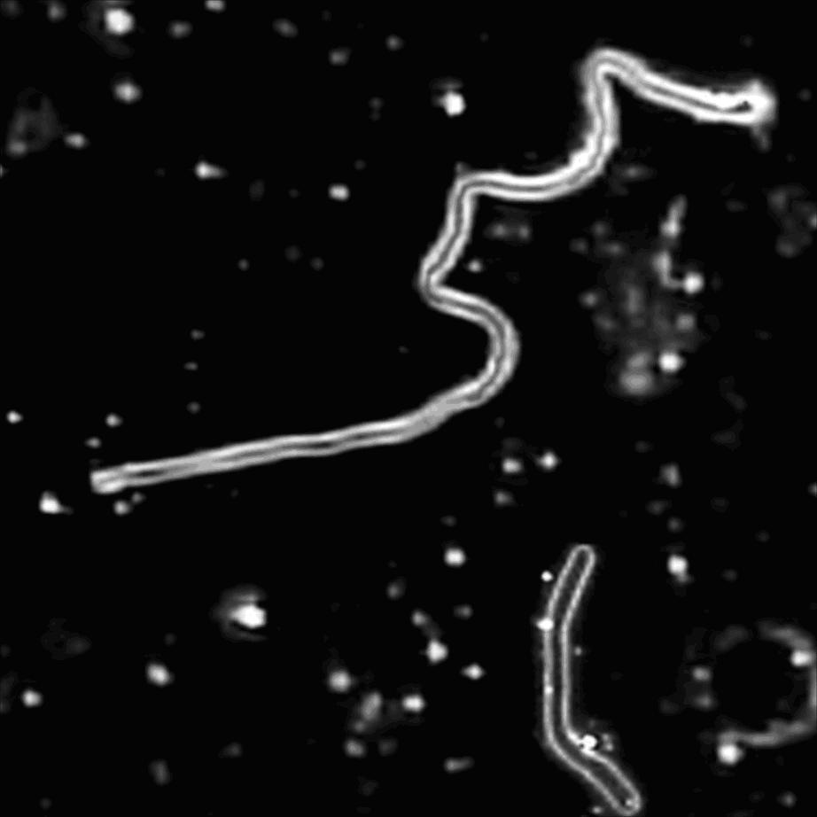
In our search for spirochetes involved in Alzheimer’s disease (13), we observed pleomorphic bacteria in the blood of healthy human subjects by dark-field microscopy.
This was a surprising finding since it is generally acknowledged that the bloodstream in healthy humans is a sterile environment (7) except when there is a breach in the integrity of the tissue membranes (6).
However, the concept of the occurrence of bacteria in the blood of healthy humans is now more plausible because of cultivation-independent laboratory approaches.
The main techniques employed in such studies include PCR amplification and sequencing of the16S ribosomal DNA (rDNA). These methods have revealed the presence of a wide diversity of microorganisms in the environment, and indeed within the human body (12).
In this report we present evidence based on molecular phylogenetic techniques and light and electron microscopy, as well as other conventional microbiological methods, for the existence of a population of bacteria in healthy human blood.
In view of the apparent controversial nature of our findings, it was encouraging to note the recent report of Nikkari et al. (14), who detected blood-associated bacterial rDNA sequences by using real-time PCR methods and a probe targeting conserved regions of bacterial 16S rDNA, and an earlier report by Tedeshi et al. (16) on the presence of pleomorphic bacteria as intraerythrocytic parasites in clinically healthy human subjects.
For light microscopic examination, blood samples from 25 healthy volunteers were drawn in a Vacutainer tube with no anticoagulants (Becton Dickinson, Franklin Lakes, N.J.); blood was drawn in the conventional manner involving antisepsis of the skin and avoidance of any introduction of external microorganisms by contamination. (Since external contamination was always a possibility, particular care and precaution were exercised at all times to avoid this.
The specific procedures, as well as appropriate controls, are specified throughout the text.)
A wet mount of the serum from the clotted blood of each sample, fresh or incubated at 30°C for between 5 to 7 days, was examined by dark-field microscopy (Leitz Dialux 20) for pleomorphic bacteria.